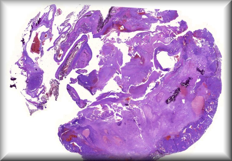
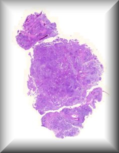
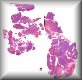
Exenteration specimen of a 70-year-old female who underwent multiple (14) surgeries for conjunctival melanoma over a period of two years.
A 65-year-old woman with a rapidly growing tumor in the upper left eyelid

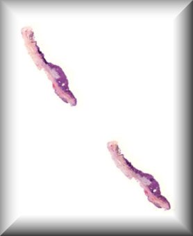
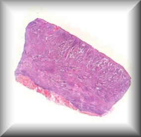
Corneal edema occurring 45 years after penetrating keratoplasty in a patient with keratoconus
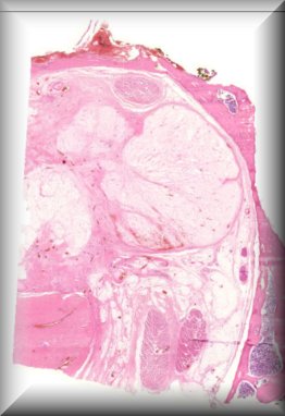
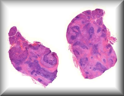
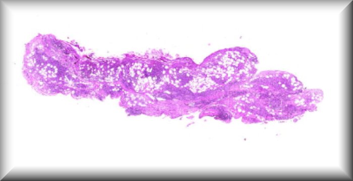
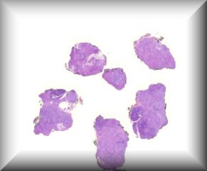
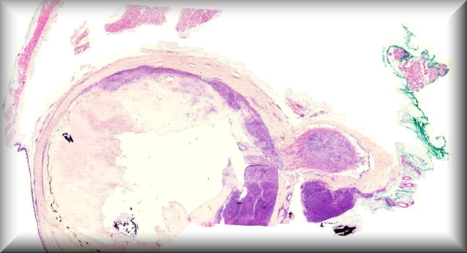
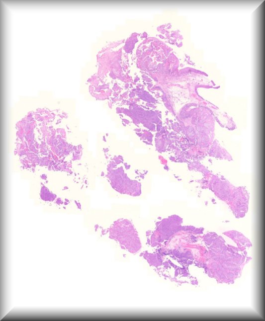
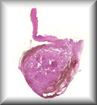
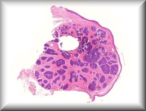
A 10-month-old boy presented with a painless scrotal mass.External examination demonstrated left scrotal non-tender hard mass measuring 4x3 cm in size, with negative transillumination test. Ultrasonography of left scrotum showed a well-circumscribed, oval shaped mass with mixed echogenicity, The mass contained both cystic and solid component, with no clear visualization of the left testicle.The patient underwent left scrotal exploration with high inguinal orchidectomy
26 year old male with history of Von Hipple Lindau syndrome. Enucleation of the left eye was performed.
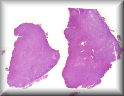
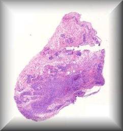
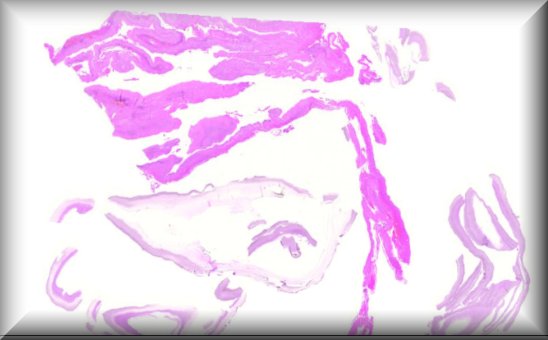
Exenteration specimen in 18y old lady after recurrence of disease.
The patient is a 64yo men with history of vision lossin the left eye (LE) in the last month due to vitreous hemorrhage. Ocular history: low vision since childhood because of choriorretinitis. Suspected intraocular tumor.
A three-year-old boy developed proptosis over three weeks. Computed tomography and magnetic resonance imaging disclosed a 3.2 x 1.9 cm soft-tissue mass of the right extraconal and intraconal orbit with sphenoid bone erosion. The tumor was debulked through an upper eyelid crease incision.
36 y old female with enlarged right lacrimal gland. Known Systemic lupus erythematosis (on Hydroxychloroquine) and anti-phospholipid syndrome. Lacrimal gland biopsied.
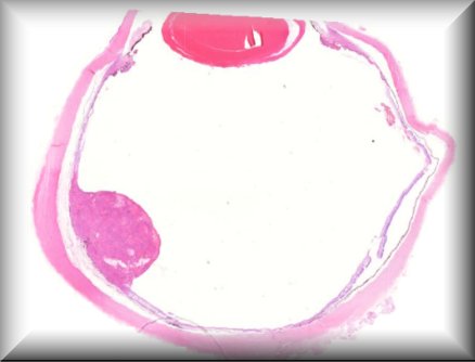
The patient was a man of 22 years who presented with ptosis and a swelling of his left upper eyelid in the temporal region. MRI of the left orbit showed a hyperintense lesion in the left upper lateral quadrant of the orbit, either arising from, or compressing the lacrimal gland. There was some displacement of the bulbus. The differential diagnosis was dermoid cyst (not entirely typical) or hemorrhagic lesion, such as hemangioma or hemorrhagis pleomorphic adenoma. The tumour was successfully excised.
71 year old woman with a presumed left upper eyelid chalazion refractory to therapy.
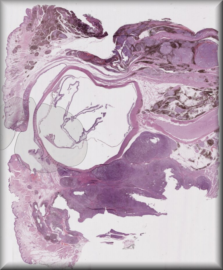
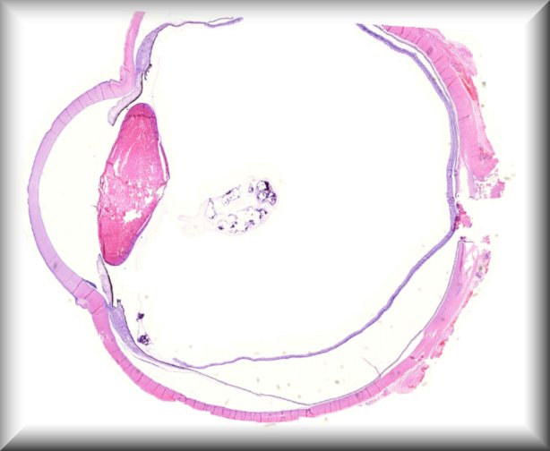
Enucleation of a painful blind eye after perforation injury 39 years ago (and multiple surgeries during the following years).
Progressive swelling of left upper and lower eyelids, conjunctival chemosis, and redness in a 47 year old female. Orbital biopsies and a further conjunctival biopsy are performed.
46 year old female. Left frontal lobe glioblastoma treated with surgery and radiotherapy. Orbital lesion for a few weeks. Two x biopsy - grossly white fish flesh appearance? Lymphoma
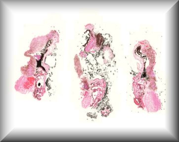
Enucleation specimen in 71 year-old man after iodium radiation.
A 68-year-old woman with a medical history of colon neoplasia treated with surgery and chemotherapy 4 years earlier with no evidence of disease in subsequent check-ups was attended the emergency department of our centre due to inflammation of the left upper eyelid of 1 month of evolution and horizontal binocular diplopia of 4-5 days of evolution. The physical examination highlighted complete ptosis of the left eye, limitation to left supra-elevation of -2 and diplopia in all positions without pain. The conjunctiva was normal coloured and cornea was transparent. There were no evidence of changes in the eye fundus. In the entire anterior orbit, in front of the orbital rim, painful stony palpation was observed, without fornix masses.
67 year old male, penetrating trauma aged 13 (tree branch), loss of light perception. Since a few months a hard subconjunctival lesion inferior on the same eye.
78-year-old male with a bulging and bleeding lesion in the caruncle of the right eye, with adhesion to the tarsal conjunctiva of the nasal third of the lower eyelid, with some keratinized areas. Resection of the lesion.
A 71-year old female patient presented with a ciliary body tumour in an eye with iris bicolor.
Breakthrough of an aggressive tumor into the orbit.
A 15 year old woman presenting with a left eye conjunctival pigmented lesion. The lesion has recently changed. Removal of the lesion has been approved. There is no particular clinical history.
45-year-old woman with swelling of the left upper eyelid for 2 months. Conjunctival biopsy.
51-years old male patient with unilateral sudden visual loss due to cellular vitreous infiltration.
http://www.pathofro.de username and password EOPS 2022
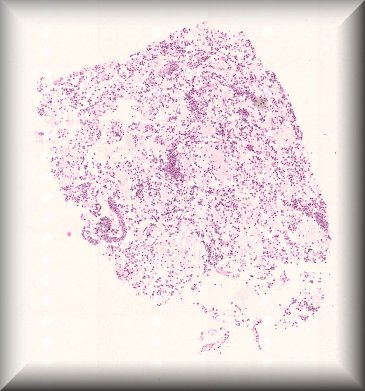
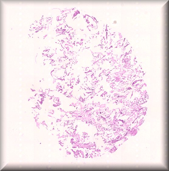
A 63-year-old patient presented with loss of vision over the course of 6 to 8 weeks. After clinical examination, there was the suspicion of uveitis under treatment with a Tyrosine kinase inhibitor. Diagnostic vitrectomy was performed.
http://www.pathofro.de username and password EOPS 2022

Intraconal mass biopsy in a 70-year-old male
77 yo male patient presenting with an upper eyelid mass. A tumor had been excised from the same area two years prior elsewhere.
An orbital malignant melanoma arising in cellular blue nevus in a 52-year-old Caucasian man.
Iridociliary melanoma in a 55-year-old woman after excisional surgery of a tumor-like lesion on her right eye iris.
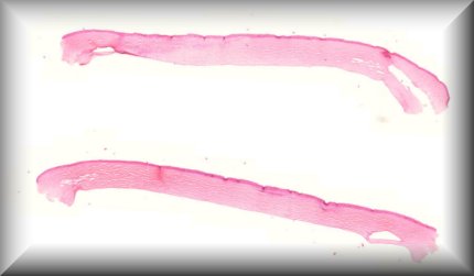
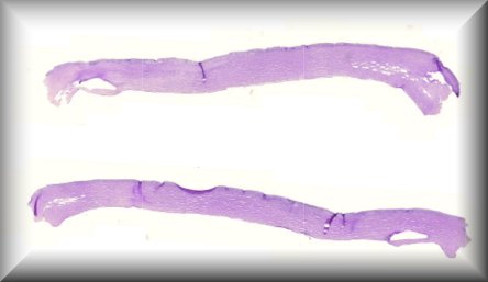
Histological findings after corneal ring segments explantation in a patient with radial keratotomy and post-LASIK ectasia.
70-year-old Caucasian male. Nodular lesion right upper lid.
Expansive lesion in the Right superomedial orbital quadrant, with grossly ovoid and lobulated contours, and 22x22x11 mm. It had regular and well-defined limits and homogeneous content, apparently contacted the eyeball, as well as the medial rectus and superior rectus muscles, deflecting them, with no signs of invasion.
A 26-year-old patient has been suffering for two years from a massive right exophthalmos.
73-year-old female with a 7mm in diameter, smooth, erythematous nodule with teleangectasias on the right upper eyelid of 4 years duration. Madarosis was also observed. Past clinical history included a previous diagnosis of breast cancer 10 months before.
Anterior chamber extension of retinoblastoma